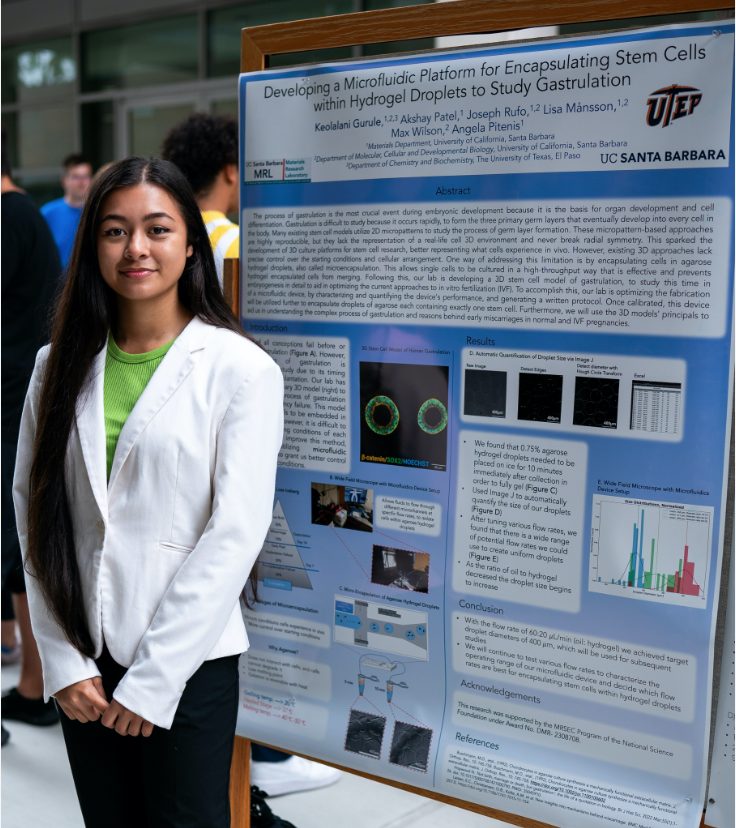
The process of gastrulation is the most crucial event during embryonic development because it is the basis for organ development and cell differentiation. Gastrulation is difficult to study because it occurs rapidly, to form the three primary germ layers that eventually develop into every cell in the body. Many existing stem cell models utilize 2D micropatterns to study the process of germ layer formation. These micropattern-based approaches are highly reproducible, but they lack the representation of a real-life cell 3D environment and never break radial symmetry. This sparked the development of 3D culture platforms for stem cell research, better representing what cells experience in vivo. However, existing 3D approaches lack precise control over the starting conditions and cellular arrangement. One way of addressing this limitation is by encapsulating cells in agarose hydrogel droplets, also called microencapsulation. This allows single cells to be cultured in a high-throughput way that is effective and prevents hydrogel encapsulated cells from merging. Following this, our lab is developing a 3D stem cell model of gastrulation, to study this time in embryogenesis in detail to aid in optimizing the current approaches to in vitro fertilization (IVF). To accomplish this, our lab is optimizing the fabrication of a microfluidic device, by characterizing and quantifying the device's performance, and generating a written protocol. Once calibrated, this device will be utilized further to encapsulate droplets of agarose each containing exactly one stem cell. Furthermore, we will use the 3D models’ principals to aid us in understanding the complex process of gastrulation and reasons behind early miscarriages in normal and IVF pregnancies.