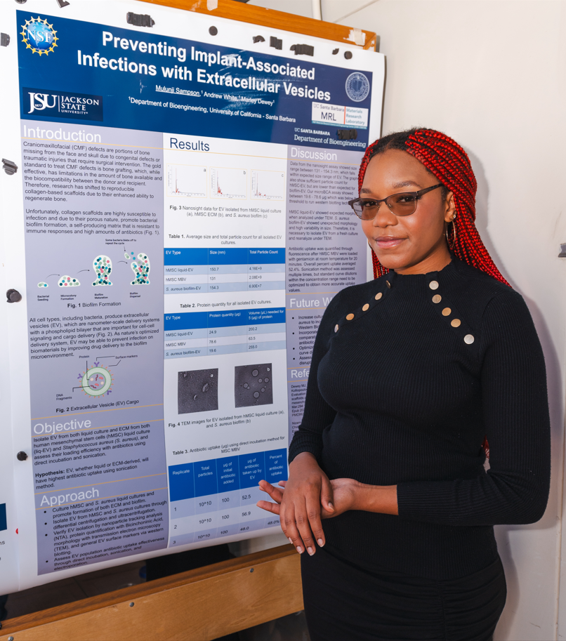
Critically-sized bone deformities in the face and skull, otherwise known as craniomaxillofacial
defects (CMF), affect hundreds of thousands of people each year worldwide. This condition,
along with defects caused by traumatic injury, is only suspected to grow with each passing year,
making medical intervention via implant more relevant by the day. With the increasing relevancy
of medical intervention surrounding CMF defects, comes increasing concern about the
biomaterials used in most medical interventions such as implants, particularly their susceptibility
to infection. This study documents human cell and infected implant material interaction after
medical intervention through extracellular vesicle (EV) loading of antibiotics. Particularly, the
capability of EVs—nanometer-sized lipid bilayer vesicles containing cargo that directly
influence cell behavior—to uptake and administer antibiotics to infected materials and human
tissue. We utilize a vast array of EV isolation techniques before approaching the process of
antibiotic loading, including ultracentrifugation, centrifugation, and the usage of size exclusion
chromatography columns. After the process of isolation is complete, we run the processed
solutions through a nanosight which would identify the size and concentration of particles within
the range of 30 to 200 nanometers. Followed by western blotting for EV protein identification,
microbicinchoninic acid assay for protein counting within an EV-isolated and particle-free
solution, and transmission electron microscopy for in vivo EV observation and modulation. We
find that after a given antibiotic is loaded into an EV through an existing method the antibiotic in
question can effectively infiltrate and eliminate the root cause of implant-associated infections,
resulting in the death of targeted microbial cells, meanwhile maintaining a safe amount of
antibiotic solution exposed to the surrounding human cells.